

Table of Contents Show
The first pregnancy scan should be a memorable experience. It’s good news to learn you are pregnant. Your 7-week ultrasound scan will give you the first glimpse of your baby. They may be a little more than a small dot on the scan, but it will be your confirmatory test to say you are pregnant.
The 7-week ultrasound will show you that your baby is about the size of a blueberry. At 7 weeks, you can determine your due date and check the heartbeat and brain development of the baby.
1. Overview of Week 7 of Pregnancy

Most people already know they’re pregnant by now. They may have missed their menstrual period, have taken a pregnancy test, or have already consulted a doctor. But, some are waiting until this week to be certain.
It’s not apparent that you are pregnant yet, but you’re noticing the signs. You may have gained weight or maybe even lost a few pounds due to morning sickness. Your breast may start getting bigger, and your pants may not be well due to bloating.
1.1 Symptoms of 7 Weeks Pregnancy
You may start to experience early pregnancy symptoms as your baby continues to grow. Here are a few symptoms you may experience during this time:
- nausea/ vomiting
- frequent urination
- fatigue
- tender and swollen breasts with darkening areolas
- mild pelvic cramps
- food aversions and cravings
- mood swings
The pregnancy hormones may make you sensitive to different odors, which can suddenly make you nauseous. Certain foods may seem more appealing to you, while your favorite food seems repulsive.
Most women experience nausea, food aversions, or cravings in their first trimester, but it can also last throughout your pregnancy. This may cause trouble for many women to maintain a balanced diet. Prenatal vitamins can bridge the gap in your limited diet.

You may want to consult your doctor if the symptoms are excessive and you cannot keep any food or liquid down for more than 24 hours.
You may also notice that you are producing excessive saliva. The cause of this symptom is unknown, but there are claims that it may be due to hormonal changes or heartburn. Be sure to keep yourself hydrated, as it can help reduce saliva production.
Fatigue is also a prominent symptom in the first and third trimesters. You may find yourself wanting to catch an extra snooze in the middle of the day. You can try some light exercise to boost your energy. But consult your doctor before you start your exercise routine.
1.2 Your Baby’s Development at 7 Weeks

As of this week, your baby is still considered an embryo and is only 1/4-inch long. The spine tail is becoming smaller and will soon disappear entirely.
The baby’s head and face are taking shape as nostrils appear and the baby’s eye lens develops. The hands and feet also start forming, but they will resemble small paddles for now.
2. Schedule your Prenatal Visit
Prenatal care appointments are essential if you haven’t already scheduled one. Most wIt will be the most thorough and lengthy visit. Omen have their first prenatal checkup this week or at week 8. During this visit, your doctor will review your medical history, do a pelvic exam, give you an estimated due date, and identify any pregnancy risks.
They will check your weight and blood pressure; and order a few blood tests, urine analysis, and even a Pap smear test. They will also do an ultrasound test to check the developing baby.
2.1 Reasons for ordering a 7-week ultrasound

There are a bunch of reasons why your doctor may recommend a 7-week ultrasound – and not all of them are dismal. The most common reason to get a 7-week ultrasound instead of at 12- or 13 weeks is to date the pregnancy accurately.
Measurements taken during an early ultrasound can determine the baby’s gestational age and accurately tell you how far along you are in your pregnancy. This can be very helpful for first-time mothers who cannot match their symptoms to pregnancy or want confirmation of pregnancy.
A dating scan or an early 7-week ultrasound is necessary for women who:
- Are unsure of their last menstrual cycle or have a history of an irregular menstrual cycle?
- Have recently had a miscarriage and conceived again.
- Having had fertility assistance or a history of obstetrics problems, a seven weeks scan can be reassuring.
- Have conceived after stopping contraception.
- Have conceived during the breastfeeding period even though they may not have resumed menstruation.
- To confirm the gestational age of the baby.
- Are experiencing blood loss after a positive pregnancy test

2.2. Other reasons your doctor may suggest an early 7-week ultrasound are:
- Confirming pregnancy with twins or multiples (especially if you’ve had fertility treatments): Multiple pregnancies include more than one baby, that is, twins, triplets, or more. In the case of twins, there could be a single gestational sac with two embryos or two gestational sacs with an embryo in each.
- Ruling out ectopic pregnancy: An ectopic pregnancy happens when an embryo implants outside of the uterus, even if it isn’t viable. If left untreated, an ectopic pregnancy may be life-threatening.
- Checking fetal heartbeat: You can hear your baby’s heartbeat in a 7-week ultrasound. This will be checked, especially when you have concerning symptoms like abdominal pain, vaginal bleeding, or spotting, which may be caused by miscarriage.
- Examine your reproductive anatomy: Complications during pregnancy may result from problems with your uterus, cervix, ovaries, or fallopian tubes.
Having a 7-week ultrasound is not a cause for alarm; it is a routine test. Sure, it could be deemed an emergency, but it could also be simply a way to collect as much information as possible to help you have a healthy pregnancy.
3. Process of Doing 7-Week Ultrasound

This may be your first pregnancy ultrasound to see your baby, so it’s bound to be an exciting time. There are two ways of doing a 7-week ultrasound – one method is transabdominal ultrasound and the other is transvaginal ultrasound. In the early weeks of pregnancy, a transvaginal ultrasound is said to provide more accurate visualization.
At 7 weeks of pregnancy, the baby may be too small to be detected on an external abdominal ultrasound. In such a case, transvaginal ultrasound is more effective as the transducer does not need to send sound waves through muscles and tissues to give returning images.
It is exciting to see your baby for the first time, but the ultrasound procedure may not be as fun. In a transvaginal ultrasound conducted at 7 weeks gestation, the lab technician will insert an ultrasound wand called a transducer into the vagina until it reaches the cervix. The technician will keep the wand in place and may maneuver it as much as needed to get a clear picture. You do not need a full bladder to do a transvaginal ultrasound1.
This form of scan is not painful, but it can be uncomfortable. The process may last longer adding to the discomfort, but your technician is trained to make you feel more comfortable, or as comfortable as you can be with an ultrasound wand inside you. There is no risk to the baby from this procedure. There is no radiation involved either. Hence, it is very safe.
Transabdominal ultrasound conducted, however, requires the patient to have a full bladder so that the uterus can be lifted out of the pelvis to allow for a more precise viewing of the embryo. As the pregnancy progresses into the second trimester, a full bladder becomes unnecessary as the uterus grows and does not fit within the pelvic rim.
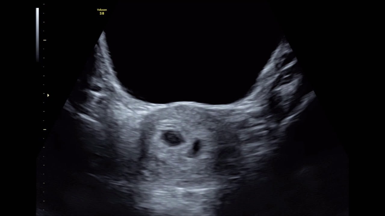
4. What will you see?
The sonographer 2will take a scan to confirm the number of gestational sacs with an embryo in the uterus and the health of the baby’s heart.
It counts the number of gestational sacs and embryos in each sac. A single pregnancy includes only one gestational sac with a single embryo inside, whereas multiple pregnancies such as twins or triplets. can contain two gestational sacs with two embryos each or two gestational sacs with an embryo each. You can use this combination to determine whether you are carrying identical or non-identical twins, or if you are expecting triplets.
You will also hear the heartbeat of the baby. The heart rate could be as fast as 110 beats per minute or more. If your baby is in a visible position, you may also see them blinking on the ultrasound.
The anatomical developments you may see on your 7-week ultrasound are:
- Gestational Sac: It is a fluid-filled space around the embryo that develops by the 5th week of pregnancy. It is a confirmation of intrauterine pregnancy. In ultrasound, it is visible as a clear, dark circular, or oval-shaped structure with white opaque contrast inside the uterus.
- Yolk sac: This is the first development inside the gestational sac, which provides nutrition and oxygen to the baby until the development of the placenta. You may spot this even before the embryo, as a small white ring inside the sac.
- Fetal pole: The first sign of your baby forming inside the yolk sac is in the form of a thick, whitish-shaped structure which may be curved or oblong depending on the duration of your pregnancy.
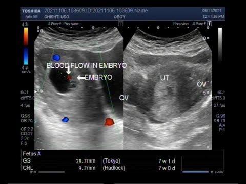
5. Measurements Taken
Your doctor will take fetal measurements of your growing baby during your 7-week ultrasound. These measurements will give your doctor the information required for the accurate dating of your pregnancy.
These measurements are used as an indicator of gestational 3age, and hence, the 7-week ultrasound is also called a dating scan.
If your embryo is visible, the technician will take a crown-to-rump measurement and the size of your gestational sac. By seven weeks, the gestational sac should reach a size of approximately 18 to 24 millimeters (mm), and your baby should be 5 to 9 millimeters (mm) in diameter.
These measurements change rapidly, with a significant difference each week as your baby is on a fast track in fetal development.

If your gestational sac measures below 18mm, your doctor may place your pregnancy a few weeks less, like maybe 5 or 6 weeks. The opposite is true if the gestational sac measures more than 24mm.
If the results of your 7-week ultrasound are not clear, your doctor may re-schedule your appointment after another one or two weeks. This happens when your doctor cannot spot fetal development clearly. You may be worried about why you cannot see a thing on your ultrasound.
In early pregnancy, your embryo or gestational sac may be just way too small to see in your first ultrasound. But don’t worry, your embryo will keep developing faster and bigger with each passing day. You may see a big difference in just a week or two.
6. Visibility of Twins
One of the main reasons for an early ultrasound scan is to check how many babies are growing in your uterus. Multiple sacs are clearly visible in this early ultrasound scan. Fraternal twins will be more clearly visible in a 7-week ultrasound. In fraternal pregnancies, two different eggs were fertilized, which creates two separate gestational sacs.
In the case of identical twins, meaning one egg was fertilized that split into two, there will be only one gestational sac. More than one yolk sac, multiple heartbeats, and fetal pole may be visible.
Sometimes, multiple gestational sacs may not be visible until later in the pregnancy, as the babies can hide behind the other sibling. Again, remember that an early ultrasound scan may not be 100% accurate.
7. Conclusion
Being 7 weeks pregnant is an exciting and essential time during your pregnancy. As the body continues to prepare nourishment for the baby’s growth, there may be some symptoms you may start to notice. They may be like nausea4, vomiting, or fatigue.
It is also an excellent time to get the first look at your baby through a 7-week ultrasound. It is an uncomfortable but harmless procedure that offers great insights into your baby’s development.
You will understand whether your pregnancy is viable, how far along you are, and how many babies you are carrying. This information is essential to plan for your pregnancy and after the coming months.
FAQ
1. What are the common symptoms at 7 weeks of pregnancy?
A: At 7 weeks of pregnancy, common symptoms may include morning sickness (nausea and vomiting), breast tenderness, frequent urination, fatigue, food cravings or aversions, mood swings, and a heightened sense of smell. However, it’s important to note that every woman’s experience may vary.
2. Is it normal to have cramping at 7 weeks pregnant?
A: Mild cramping can be normal during early pregnancy as the uterus expands and the ligaments stretch. However, severe or persistent cramping, accompanied by heavy bleeding or other concerning symptoms, should be reported to a healthcare provider.
3. How big is the fetus at 7 weeks pregnant?
A: At 7 weeks, the fetus is approximately 0.51 inches (1.3 centimeters) long, about the size of a blueberry.
Read more
- Liu, Linly, et al. “Diagnostic accuracy of transvaginal ultrasound and magnetic resonance imaging for adenomyosis: systematic review and meta‐analysis and review of sonographic diagnostic criteria.” Journal of ultrasound in medicine 40.11 (2021): 2289-2306. ↩︎
- Marriner, Mieko. “Sonographer quality management.” Journal of Echocardiography 18 (2020): 44-46. ↩︎
- Lende, Michelle, and Asha Rijhsinghani. “Gestational diabetes: overview with emphasis on medical management.” International journal of environmental research and public health 17.24 (2020): 9573. ↩︎
- Gan, Tong J., et al. “Fourth consensus guidelines for the management of postoperative nausea and vomiting.” Anesthesia & Analgesia 131.2 (2020): 411-448. ↩︎
Last Updated on by Sathi Chakraborty, MSc Biology


