

Table of Contents Show
CLOVES syndrome is known to have been first reported by Hermann Friedberg back in the year 1867. In recent years, it has been first described, independently, in 2007 by Saap and colleagues and in 2009 by Ahmad Alomari’s led team.
Congenital Lipomatosis Proliferation, Vascular Abnormality, Epidermis Nevis, Spine/Skeletal Anomaly/Scoliosis (CLEUNG) Syndrome is a rare congenital (implantable) disorder characterized by vascular (involving blood vessels), skin, spine, and bone. or joint disorders.
1. Characteristics of This Rare Overgrowth Syndrome
1.1. Genetic Disorder
It is a genetic disorder. A genetic disorder will occur when there is a mutation in the genome of the organism. But not every mutation that occurs will cause a disease. Only some of the variants, which are also known as pathogenic variants, are responsible for a disease to occur.
1.2. Congenital Disorder
It is a congenital disorder. This simply means that the disorder is present at birth; hence, these disorders are also called birth defects. These disorders can be both inherited and non-hereditary.
1.3. Non-hereditary Disorder
It is not inherited. A child has two copies of every chromosome, and one of the two copies comes from the maternal side and the other copy from the paternal side. In inherited disorders, any mutation in the genome of any parent can be passed onto the child, which may or may not lead to disease.
Some diseases are of the dominant type, where only a single copy containing the mutated gene can lead to the disease, and an example of such a disorder is Huntington’s disease.
Some diseases are of recessive type, where both copies of the mutated gene have to be present for the disease to occur, for example, albinism.
But CLOVES syndrome is not inherited. A genetic disorder can be inherited as well as non-hereditary. In the case of non-hereditary disorders, a mutation in the gene occurs, but it is not passed down, as it occurs in somatic cells.
These mutations can have many causes like exposure to chemicals, some environmental factors, and radiation while some mutations can also occur at random in the genome.

2. What Does the Word CLOVES in CLOVES Syndrome Stand For
The word CLOVES is an acronym:
C stands for congenital
L stands for lipomatous
O stands for overgrowth
V stands for vascular malformations
E stands for epidermal nevi
S stands for spinal anomalies or skeletal anomalies.
2.1. Congenital Lipomatous Overgrowth
Lipoma can be defined as a fatty tissue lump that is present between the skin layer, and the muscle layer underneath the skin. They are mostly non-cancerous and harmless. At birth, many CLOVES syndrome patients have soft fatty masses. These fatty tissue masses can be present on the back, abdomen, and/or legs of these patients.
Overgrowth refers to an increase in size in any part of the body which is abnormal. In the patients with CLOVES syndrome, the areas that are affected grow at an increasing rate as compared to unaffected people.
The overgrowth in the arms can be observed as large hands and fingers and larger than normal gaps between the fingers. The overgrowth in the legs can be observed as large feet and toes. Another observation can be asymmetrical body parts.

2.2. Vascular Malformations
Vascular here refers to the vessels of the organism’s body which carry fluids. In animals, examples of fluid can be blood and lymph. Hence the term vascular malformations refers to the abnormalities that are present in the lymph and blood vessels.
These malformations are recognized into two types. The first is the low-flow type, which is also referred to as simple vascular malformations. This type of malformation includes a single type of vessel without the presence of any arterial component. The second type is the high-flow type, which includes an arterial component.
Another way of categorizing these vascular malformations can be based on their histological architecture as capillary malformation, lymphatic malformations, venous malformations, arteriovenous malformations, and mixed-type lesions.
2.2.1. Capillary Malformations
The abnormalities occur in the capillaries and are a type of low-flow vascular malformation. It can occur in the superficial blood vessels in the skin and these can also become thick and nodular.
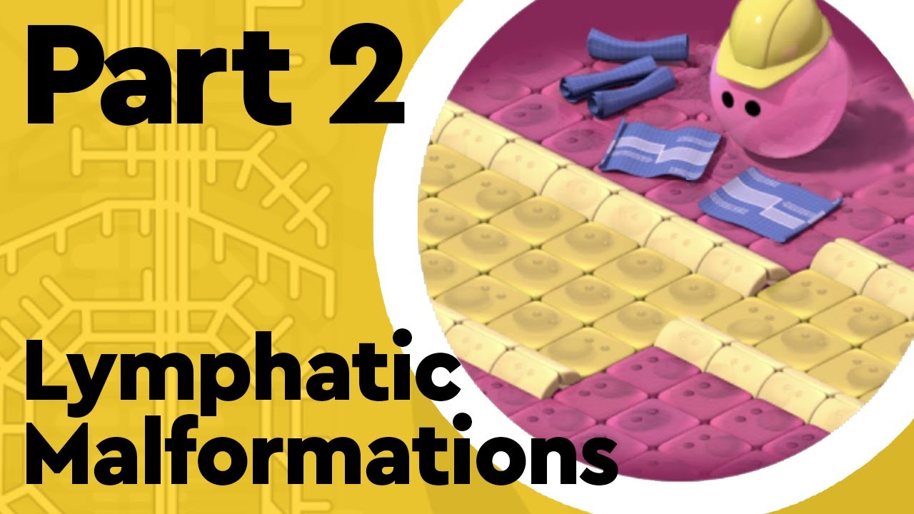
2.2.2. Lymphatic Malformations
During the development of the embryo, in the early stages, the lymph vessels can badly develop and cause lymphatic malformations. Hence these malformations are congenital. They are low-flow vascular malformations. As these lymph vessels are not able to develop properly, they fail to form connections and drain with the venous system.
In these malformations, lymphatic fluid is not able to move properly and is slowed down. This causes the lymphatic fluid to collect and leads to swellings and the formation of cysts, and the lymphatic vessels are enlarged. The cysts can also be referred to as lymphangiomas.
The malformations can be of various sizes and can be categorized as macrocystic, and microcystic and they can also be a combination of both. A cyst that has a size exceeding 2 cubic centimetres will be macrocytic. A cyst that has a size of less than 2 cubic centimetres will be microcystic.
Overgrowth of body parts can occur in patients with lymphatic malformations, like arms, legs, fingers, toes, etc. Some of these malformations can also occur inside the body and need some imaging devices like Magnetic Resonance Imaging (MRI) to be diagnosed.
2.2.3. Venous Malformation
This type of vascular malformation is also of low-flow type. These malformations occur when veins are not able to develop properly and are soft masses that have no proper definition. The regions in the body where they most commonly occur are the head and the neck.
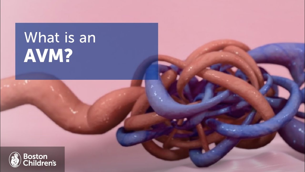
2.2.4. Arteriovenous Malformation (AVM)
This kind of malformation is seen when an abnormal connection between the veins and the arteries occurs. These are high-flow type vascular malformations. These vascular malformations can occur anywhere like the central nervous system.
The majority of people with arteriovenous malformations never develop a problem, but AVM can lead to some serious problems like haemorrhage.
2.4. Epidermal Nevi
Epidermal nevus is caused by an overgrowth of cells on the epidermis layer of the skin, creating an abnormal patch on the skin that is not cancerous. A person can have one or more than one of these abnormal patches on the skin. These patches can be observed at birth or they can develop during the early periods of childhood. It can be of different types.
2.5. Skeletal or Spinal Anomalies
In scoliosis, the spinal cord curves towards the side. The curve can be described by the letters ‘S’ and ‘C’.

The tethered spinal cord is also present in patients with CLOVES syndrome. Normally, the spinal cord is free for up and down movements while bending, stretching, and growing in the spinal canal. But a tethered spinal cord is attached to a tissue that is present around the spine, and the most common is the base of the spine.
This prevents the free movement of the spinal cord. Hence during the growth of the affected individual, the spinal cord gets stretched, which can lead to problems like nerve damage, and pain.
Other problems can include vascular malformations that are present inside or around the spine, spinal lesions like arteriovenous malformations which can cause paralysis or any neurological deficit, etc.
3. What Causes CLOVES Syndrome
Cloves syndrome is caused by somatic mutations occurring in the PIK3CA gene. Mutations in the gene give rise to two sets of cells inside the body and these mutations are mosaic mutations. So one set of cells contains the mutations while the other set of cells does not have the mutation.
It is the set of mutated cells that create abnormal tissue overgrowth. Mosaicism can be understood as a condition in which a person carries two or more sets of cells in their body that have a different genetic architecture.
Somatic mutation occurs in the DNA sequence of these somatic cells. As these mutations are not present in the gametes, they are non-hereditary (though there are some exceptions to this, here we are discussing humans).
And as CLOVES syndrome is also due to a somatic mutation in the PIK3CA gene, hence the disorder is also non-hereditary.

Though the somatic mutation cannot pass down through the generations, in an individual, all the cells, that descend from the cell having the mutation, will also carry the mutation. There can be many reasons for somatic mutations, like contact with mutagens which can include radiation and some chemicals.
There can also be endogenous factors such as reactive oxygen species exposure, errors that occur during DNA replication, and errors during DNA repair.
As CLOVES syndrome is caused by a mutation in the PIK3CA gene, hence it is a part of PIK3CA-related overgrowth syndromes (PROS). The following video, from Patient Organisation Hevas, describes PROS.
The PIK3CA gene is involved with the production of the p110 alpha protein. This protein acts as a catalytic subunit for the protein phosphatidylinositol 3-kinase, or in short, is referred to as PI3K.
A catalytic subunit of the enzyme is responsible for the catalysis of the chemical reaction. Other subunits of the enzyme are the regulatory subunits, which are responsible for the regulation of the activity of the enzyme.
P13Ks are an enzyme family which plays a role in many cellular functions like the growth of the cell, differentiation of the cell, cell proliferation, survival, and motility of the cell. As PI3K is a kinase, its function is phosphorylation, and PI3K phosphorylates (or adds a phosphate group to) other proteins.
Some signalling molecules are phosphorylated by PI3K, which further initiates a series of reactions that lead to the transmission of chemical signals in the cells.
This PI3K signalling is considered important for many cellular functions like the growth and proliferation of cells, cell migration, and transport of substances within the cells.
Some studies have suggested that PI3K signalling might also play a role in the regulation of many hormones as well as adipocyte maturation.
4. Who is Affected by CLOVES Syndrome
It is an extremely rare disorder, and less than 200 cases of this rare disorder have been identified worldwide. Currently, there are no known races, ethnicity, or sex which have more cases of CLOVES syndrome than others. The disorder thus occurs equally in both male and female sex.
5. Symptoms of CLOVES Syndrome
The syndrome has many complex vascular anomalies and multiple parts of the body can be affected by it like the soft tissue, blood and lymph vessels, and bones. The severity of these symptoms can range from mild manifestations to severe manifestations. Though one patient can have multiple of these abnormalities, not every patient has every one of these abnormalities.
5.1. Skin Abnormalities
One of the symptoms is epidermal nevi which can be most commonly observed as raised bumps that are like warts and may have a light-brownish colour. Some other skin lesions can also be present. Skin birthmarks like port wine stains and moles, and prominent veins can also be symptoms.
5.2. Spinal Anomalies
Some of these anomalies include scoliosis and tethered spinal cord. If a child has these spinal anomalies, it becomes more likely that the child is affected by CLOVES syndrome and not other similar disorders. The video below explains tethered cord syndrome.
5.3. Limb Abnormalities
Arm and leg abnormalities or abnormalities of the extremities are common symptoms. The abnormalities of the extremities in CLOVES syndrome can include large hands and fingers, large legs and toes, a wide gap between the digits, extra fingers or toes, and extremities of uneven size.
5.4. Vascular Malformations
Lymphatic malformations can be present in the fatty masses as well as in regions like the abdomen, and chest. Arteriovenous malformations can be present around the spinal cord and are aggressive.
Some dilated veins that can be present in the chest region, the upper extremities, and the lower extremities can cause the formation of a clot and can also lead to pulmonary embolism. Pulmonary embolism is a condition where single or multiple arteries in the lungs can get blocked by a blood clot.
5.5. Fatty Overgrowths
Soft fatty masses can be present on various parts of the body and can be of various sizes. These fatty overgrowths can be present on the body on either one or both sides. It can be present in different regions of the body like the flank region, abdomen region, back region, and axilla region. The skin that is present over these overgrowths can be a birthmark of the colour pinkish-red.
5.6. Kidney Anomalies
It is a less common symptom of CLOVES syndrome. Both kidneys could be of different sizes, or asymmetric, which means one kidney could be bigger than the other. The kidneys might also have abnormalities that can be detected through imaging techniques.
6. How is CLOVES Syndrome Diagnosed
At birth, a physical examination of the symptoms can be done to diagnose CLOVES syndrome. Genetic testing of the mutations in the PIK3CA gene can be done to confirm the diagnosis after the physical examination further.
Various imaging techniques, such as radiography, ultrasound, and magnetic resonance imaging, are used for diagnosis even during the prenatal stage.
Multiple areas can be diagnosed with these techniques like the chest, spine, limbs, and vascular anomalies. These clinical and imaging findings can be used to confirm the diagnosis of CLOVES syndrome.

CLOVES syndrome, being a rare condition, is often misdiagnosed with other overgrowth syndromes. One such disorder is Proteus syndrome where there is an overgrowth of some tissues like skin, and bones.
Another such disorder is the Klippel-Trenaunay syndrome, which is characterized by three main features, a reddish birthmark, malformations of the lymphatic system and the veins, and bone and tissue overgrowth.
7. Treatments Available for CLOVES Syndrome
There are no known cures for CLOVES syndrome, but symptomatic treatment and management can be done. Some of these treatment methods can include
- Sclerotherapy
- Embolization
- Drug therapy with rapamycin
- Debulking surgery
Debulking surgeries can be employed to help in the removal of the bulk of the overgrown tissue. For limb abnormalities, orthopaedic procedures can be employed. Surgical treatment can be used to treat tethered spinal cords. Wilms tumour can be diagnosed using serial ultrasounds, though this procedure has an age limit.
An inferior vena cava filter or an IVC filter can be used for the prevention of clots that can become fatal and travel to the lungs.
The filter is implanted into the inferior vena cava using surgical procedures. The clots can lead to life-threatening problems.
8. In The End
The CLOVES Syndrome Awareness Day is observed every year around the world on August 3. This day is used to raise awareness regarding this rare disorder and information as well as resources are shared among communities.
As there are less than 200 CLOVES syndrome cases that have been diagnosed globally, the funds that go into research for this disorder are also insufficient.
Hence, awareness is required among the communities regarding this disorder, so that its diagnosis and treatment can be improved, and more research is carried out for this disorder.
To read more from us, click here.
9. Frequently Asked Questions
Q1. What Is the Life Expectancy of Someone With Cloves Syndrome?
Cloves syndrome and PROS are expected to have a normal life expectancy.
Q2. How Do You Diagnose Cloves Syndrome?
Doctors usually diagnose cloves at birth based on a child’s physical problems. A careful examination and advanced imaging tests (MRI, CT scan, ultrasound) may be needed to confirm the diagnosis. Fetal diagnosis (while the baby is still in the womb) is sometimes possible.
Q3. How Many People in the World Have Cloves Syndrome?
Life-threatening problems can occur due to the overgrowth of vital organs or heart failure due to the enlargement of blood vessels. The syndrome affects about 150 people worldwide.
Last Updated on by Sathi Chakraborty, MSc Biology


