

Table of Contents Show
Have you ever wondered why a regular gynaecological check-up is needed?
Often we tend to ignore the campaigns persuading us about a regular check-up considering it an unnecessary expenditure of both money and time, but in reality, it reveals many of our underlying symptoms of diseases or any tumor or cyst that was silently paving its way in the body.
Gartner’s duct cyst1 is one such condition that tends to affect many women but can be easily treated as well.
A. Gartner’s duct cyst development
During the embryological development in both sexes, there are two kinds of ducts, the Wolffian ducts 2(mesonephric) and the Müllerian ducts 3(paramesonephric).
The internal urogenital tract is derived from both of these ducts. But in the upcoming weeks of pregnancy, they differentiate into two different tracts in both males and females.
In the case of females, during the eighth week of pregnancy, the Müllerian ducts after fusing distally develop into the uterus, fallopian tubes, cervix, and upper vagina while the Wolffian ducts gradually regress.
But if the Wolffian duct fails to regress, then the ducts in vestigial form can lead to vaginal inclusions like Gartner duct cyst. Gartner cyst is also called a benign vaginal cyst4.
B. Location of true Gartner’s duct cysts
Often Gartner’s cysts are confused with Bartholin’s gland cysts and other vaginal ducts but there are some slight differences in their location.
A true Gartner duct cyst is located at the right anterolateral vaginal wall (mostly). Often it is found in the proximal third of the vagina. Sometimes it may also develop in the vulva as vulval Gartner cysts. So, considering all factors, the most common location is the vaginal wall.
The remnants of the Gartner ducts sometimes secrete fluid and develop in cysts. These generally occur in the late adolescence stage. Although vaginal cysts are quite rare in newborns, sometimes in very few cases, they might have neonatal Gartner duct cysts.
C. Complications associated with Gartner’s Duct Cyst-
In obstetrics and gynecology, health issues are associated with a number of other complications. Improper evolution of the Wolffian duct in the embryonic stages and persistent Gartner duct cyst can lead to other serious health complications like-
- Ectopic Ureter
- Unilateral renal dysgenesis
- Renal hypoplasia
- Congenital uriogenital anomalies
- Abnormalities in the metanephric urinary system.
All the above complications have often been reported along with Gartner Duct Cyst. These are generally asymptomatic and may be discovered during a routine gynecologic examination.
Sometimes some mild symptoms can be observed like-
- Infections in the Reproductive system.
- Vaginal Discharge.
- Urinary incontinence and other urinary symptoms.
- Pain in the abdominal region.
- Improper bladder functioning.
- Pelvic pain
- Urinary retention
- Prolapsed urethra
- Bladder dysfunction
- Congenital urogenital anomalies
- Ipsilateral renal dysgenesis

Once in a case report, it was found that a large mesonephric cyst resulted in severe pelvic pain in a teenage girl due to acute adnexal torsion.
In another case report, Gartner duct cysts were also associated with the single vaginal ectopic ureter.
So, if any of the symptoms arise, one should get a checkup or pelvic examination as there might be some vaginal cysts like the Gartner duct cysts.
D. Differential diagnosis of Gartner duct cyst
There are many kinds of vaginal wall inclusion cysts like the Bartholin gland cyst, Gartner’s cyst, benign cystic lesions, and others.
Careful analysis and various tests help in a better conclusion of the type of cyst and the following treatment procedures.
The differential diagnosis of Gartner Duct cyst includes-
- Bartholin gland cyst
- Uterine prolapse
- Cystocele
- Rectocele
- Urethral diverticulum
- Endometriosis
- Malignant growth
- Epidermal inclusion cyst
- Benign gynecologic lesions
- Ipsilateral renal agenesis
Difference between Bartholin gland cyst and Gartner’s duct cyst-
In the case of the Bartholin gland cyst, it is mostly found in the posterolateral wall of the vagina and is associated with the Labia majora5, but the Gartner duct is found mainly in the anterolateral wall of the vagina mainly on the right side.
Uterine prolapse occurs due to the loosening of uterine ligaments leads to the drooping of the uterus and can lead to pelvic pressure due to prolapsed uterus. In the case of a large Gartner cyst, some can also experience the same pressure in the pelvic region.
E. Occurrence of Gartner Duct cyst and other vaginal cysts in the female population
Studies and statistics in the field of obstetrics and gynecology have revealed that vaginal inclusion cysts are present in almost 1%-2% of the entire female population while 10% of all benign cysts are Gartner cysts.
Gartner’s duct cyst is a type of vaginal inclusion cyst. Gartner ducts are present in almost 25% of all adult women and in only 1% of women, it develops into vaginal Gartner’s cyst.
F. Diagnosis of the Gartner’s duct cyst
During female pelvis checkups, the presence of Gartner’s duct cyst can be observed. In most of the patients, severe symptoms are not observed, it is found accidentally.
Ways to confirm the Gartner’s duct cyst are:
-
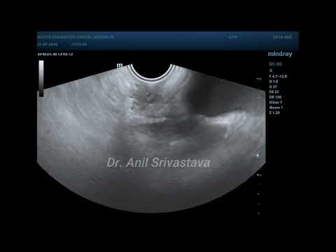
Transvaginal ultrasound
It is the technical process by which Gartner cyst dust is commonly diagnosed.
It can be used to rule out the urethral diverticulum as well. It is a rare condition in which an unwanted pocket develops from the urethra. It occurs most often in females.
Transvaginal ultrasound is the most common and cost-effective noninvasive process.
-
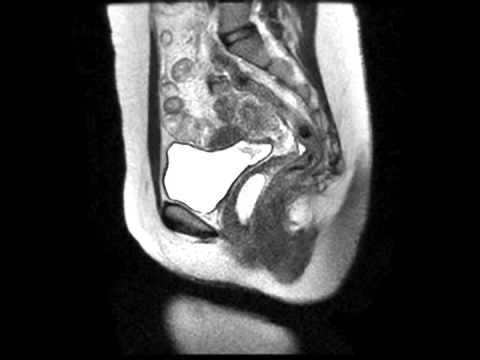
Magnetic Resonance Imaging
Although it may be done for a clarified conclusion, it does not add up much after the transvaginal ultrasound.
Moreover, it is quite expensive and hence often not recommended.
-
Speculum Examination
It is the process in which through a certain device the vagina and cervix are observed. This method is often used to detect vaginal Gartner cysts.
-
Provisional diagnostic methods
Sometimes provisional diagnostic methods are also employed like blood sample analysis, urinalysis, diagnostic laparoscopy, the study of pelvic anatomy, pregnancy tests, and transabdominal ultrasound to check the uterus, fallopian tubes, and cervix.
All these can be performed to know if there are any anomalies in the genitourinary and abdominal systems.

G. Gartner’s duct cyst and malignant transformation
Whenever there occurs any cysts or tumours, the first and foremost question that arises in our minds is if there are any chances for malignant transformation. For most of the cysts, the chances are quite less, but however, we still should not undermine their chances.
The best method to reduce its chances is to do regular checkups. Regular checkups will let one know if the once benign lesions have turned malignant in any way which then shall require immediate surgical intervention.
In the case of Gartner’s duct cyst, the chances for malignant transformation are quite less. But if there are drastic changes in size, and texture the best option will be surgical removal. Surgical excision will ensure the complete removal of the malignant mass.
Moreover, certain histopathological studies may also be done to confirm the results which may reveal that the boundaries of the cyst may be surrounded by cuboidal epithelium. Smooth muscles may also be found along with the cyst.
Hence, in certain cases, surgical management remains the most suitable choice.

H. Size range of Gartner’s duct cysts
Sometimes one may feel any slight outgrowth on the vaginal wall and tend to ignore it until and unless it causes any pain or obstetrical problems. If the size increases then one may feel it or have problems like sometimes vaginal discharge or even hindrance in tampon insertion.
In most cases, the size of Gartner’s duct cyst is less than 2cm in diameter and is found in the submucosal layer. But in certain the cyst can become a large Gartner cyst by attaining a size of up to 4.1cm along the diameter. In one case report, a Gartner’s duct cyst up to 6 cm in size was found.
I. The treatment procedure for Gartner’s duct cysts
As most of the patients are asymptomatic hence it is quite difficult to early diagnose Gartner’s duct cysts.
In the initial stages, if the size is quite small, it does no harm and does not even create any major obstetrical complications. So the doctors prefer to keep it without any surgical management but under regular inspection. This is because this gynecologic surgery is quite complicated and hence often not recommended.
In the case of symptomatic patients, the treatment procedures mostly followed in clinical practice include-
- Cyst drainage
- Injection and aspiration
- Inta-cystic tetracycline
In the case of more severe patients, the best way out is surgical excision. Cyst marsupialization may also be done depending on the cases. It is a minimally invasive process.

Whatever may be the severity of the cyst, one must be under a long-term check-up. This needs to be done to prevent any recurrence of any other cysts in the female pelvis and reproductive tract. One should keep in contact with the doctor and never discontinue their regular checkups.
Therefore, in conclusion, although Gartner’s duct cysts are generally not much life-threatening but taking medical consultation and being under medical supervision is the safest option.
No woman should neglect or hesitate about any health issues related to obstetrics and gynecology.
Many women even today, due to social myths and taboos hesitate to consult any professional doctors, but this only aids in worsening their health conditions. The negligence of today may lead to serious health issues in the coming days. The sooner we realize its vitality, the better treatment and healthy life we get in return.

- Letizia, Matthew J., and Joseph VM Kelly. “Case report: Gartner’s duct cyst.” Emergency Medicine News 33.5 (2011): 35. ↩︎
- Hannema, Sabine E., and Ieuan A. Hughes. “Regulation of Wolffian duct development.” Hormone research 67.3 (2007): 142-151. ↩︎
- Chandler, TM1, et al. “Müllerian duct anomalies: from diagnosis to intervention.” The British journal of radiology 82.984 (2009): 1034-1042. ↩︎
- Eilber, Karyn Schlunt, and Shlomo Raz. “Benign cystic lesions of the vagina: a literature review.” The Journal of urology 170.3 (2003): 717-722. ↩︎
- Hunter, John G. “Labia minora, labia majora, and clitoral hood alteration: experience-based recommendations.” Aesthetic Surgery Journal 36.1 (2016): 71-79. ↩︎
Last Updated on by ayeshayusuf


